This is one in a series of articles that provide detailed and updated information about Dental Bone Graft.
In this specific article, which focuses on Dental Bone Graft – Applications, you can read about:
For additional articles about Dental Bone Graft, see the Topic Menu.
Dental bone graft for the lower jaw
Resorption of alveolar bone is commonly seen in the posterior part of the alveolar ridge. This can cause patients to bring their jaw forward while chewing, leading to an incorrect chewing habit. To avoid this, it is advised to replace missing teeth, and if there is insufficient bone, a bone graft should be used.
The procedure for bone graft placement involves the following steps:
- Identifying the appropriate site for bone graft placement.
- Using local anesthesia to numb the site in preparation for the bone graft surgery.
- Making a small incision in the gums and retracting the gum tissue slightly for visualization of the alveolar bone.
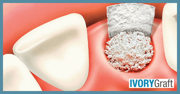
- Cleaning and disinfecting the area of defect on the alveolar bone thoroughly and placing the bone grafting material onto it.
- Typically, placing a separating membrane between the gum tissue and the bone graft is helpful to protect the graft and provide more space for bone regeneration.
- Repositioning the gum tissue back onto the membrane and placing stitches to close the incision.
After the surgery, patients are given post-operative instructions, which include managing anticipated pain, swelling, and bruising that may last for a few days. Prescriptions for antibiotics and painkillers are provided to prevent infections and alleviate pain symptoms.
Dental bone graft for the upper jaw
A dental bone graft for the upper jaw is a procedure similar to that for the lower jaw. It involves adding bone material to the upper jawbone to increase bone density and volume.
The reasons for needing a dental bone graft in the upper jaw are similar to those in the lower jaw. Common reasons include:
- Preparing the jawbone for dental implants: A bone graft may be necessary to ensure there is sufficient bone to support the implant securely.
- Providing support for a denture: A bone graft can help create a stable base for dentures, improving their fit and function.
- Addressing bone loss due to trauma or other factors: A bone graft may be used to restore lost bone in the upper jaw due to injury, gum disease, or tooth extraction.
The procedure for a dental bone graft in the upper jaw is generally the same as for the lower jaw, involving identification of the appropriate site, anesthesia, incision, cleaning and disinfecting the bone defect area, placing the bone graft material, using a separating membrane, repositioning the gum tissue, and closing the incision with stitches.
Post-operative instructions and prescriptions will also be provided to the patient to manage pain, swelling, and potential infections.
Dental bone graft for deep pocket
A healthy sulcus is 0-3 mm deep and can be easily cleaned with a toothbrush and floss. However, when the depth exceeds 3 mm, it becomes nearly impossible to maintain cleanliness at home using routine oral hygiene methods. In such cases, periodontal surgery or osseous surgery is performed for pocket depth reduction or pocket correction. After the surgery, it becomes easier for patients to maintain oral hygiene at home.
During the process, the periodontal surgeon makes an incision in the gums where pockets have formed due to bone loss. The gums are then moved away from the teeth and bone to access the tooth roots and bone. The roots are thoroughly cleaned to remove plaque or calculus and disinfected. The bone in that area is checked for irregularities; if any are found, the bone is smoothed to help reduce pocketing.
After smoothing the bone, the graft is placed in areas where there is a gap between the tooth root and bone, or between the roots of the teeth. The bone graft stimulates bone-forming cells (osteoblasts) to regenerate new bone, increasing the height and width of the bone around the tooth and providing better stability.
The most critical aspect of periodontal surgery is managing the healing phase because bone and gum tissue heal at different rates. Gum tissue heals much faster than bone, so a membrane barrier is placed between the bone and gum tissue to keep the new bone in place during healing. This prevents gum tissue from filling the space where the bone was lost, allowing new bone to grow where needed. Both the bone graft and membrane are absorbed and replaced with new bone. This technique is called Guided Tissue Regeneration (GTR).
Dental bone graft for wisdom teeth
After a wisdom tooth removal, there can be significant bone loss, the extent of which may vary based on the amount of bone removed during the impaction surgery. A bone graft can help preserve the bone in the extraction site and prevent deterioration.
The following steps are involved in the process:
- Extraction of the impacted wisdom tooth.
- Removal of any damaged or diseased tissue from the area.
- Placement of the bone graft into the socket of the extracted tooth.
- Covering the area with a protective membrane or collagen sponge.
The bone graft material fuses with the existing bone in the area, creating a solid foundation for future dental work. The healing process for a dental bone graft in the area of a wisdom tooth extraction typically takes several months.
After the procedure, patients may experience some swelling, discomfort, and minor bleeding for a few days. Pain medication may be prescribed to manage any discomfort. Patients will need to follow specific aftercare instructions, including avoiding certain foods and activities, to promote healing and prevent complications.
Dental bone graft after extraction
ooth extraction is one of the most common dental procedures. After tooth extraction, the normal pattern of bone healing is resorption, which results in hard and soft tissue defects in the alveolar bone. Loss of hard and soft tissue in the jaw directly affects the functional and aesthetic composition of the oral and maxillofacial region. Thus, these changes leading to a reduction in the quantity and quality of bone can compromise the functional and aesthetic outcomes of both implants and fixed bridge restorations.
The loss of vertical ridge height is greater compared to the horizontal ridge. These alveolar bone changes result in thin bone volume, which often compromises implant placement. Bone grafting after extraction can be done to prevent hard and soft tissue loss. Many studies have shown that residual ridge resorption is less in cases where a bone graft is placed after tooth extraction, as compared to cases where no graft is used.
The main purpose of a bone graft after extraction is to correct bony defects and preserve tissue structures for subsequent tooth replacement therapy. Therefore, proper planning before extraction is recommended, which includes identifying the correct site for placement.
Tooth socket classification when the tooth is still present:
- Type 1 socket—Buccal plate present and soft tissue present:
- Type 1a socket: thick biotype, posterior tooth, and buccal plate present: no graft needed.
- Type 1b socket: thick biotype, anterior tooth, and buccal plate present: clot stabilizer needed.
- Type 1c socket: thin biotype, anterior or posterior, and buccal plate present: bone graft needed.
- Type 2 socket: Buccal plate missing, but soft tissue present: bone graft +/- membrane (if graft containment is needed).
- Type 3 socket: Buccal plate missing and soft tissue missing: bone graft + membrane +/- biologic agent (consider soft-tissue graft if keratinized tissue is less than 2 mm).
Dental bone graft without extraction
A dental bone graft can sometimes be performed without extraction, typically to address a specific area of bone loss or damage for reasons other than extraction. This type of bone graft is called ridge augmentation.
If a patient has experienced bone loss due to periodontal disease, trauma, or other factors, ridge augmentation may be recommended. This procedure involves adding bone material to a specific area of the jawbone to create a more solid foundation for a dental implant or other dental work.
During a ridge augmentation or ridge preservation procedure, a small incision is made in the gum to expose the area of the jawbone where resorption is present. Bone graft material is then added to the area and secured in place by stitching the incision closed. A protective membrane or collagen sponge may also be placed over the graft site to further promote proper bone healing.
After the procedure, patients are asked to follow specific aftercare instructions. Pain medication may be prescribed to manage any discomfort. For a few days after the augmentation procedure, patients may experience some swelling, discomfort, and minor bleeding.
The healing process for a ridge augmentation procedure typically takes several months, during which time the bone graft material will fuse with the existing bone.
Dental bone graft for gum disease
If proper oral hygiene is not maintained, bacteria in dental plaque can invade deep into the gums, causing inflammation and tenderness. In the initial stage of gum disease (gingivitis), swelling and bleeding of the gums may occur. If left untreated, gingivitis can progress into the most advanced form of gum disease, called periodontitis. Untreated gum disease can lead to tooth loss, gum tissue loss, and eventually bone loss.
Signs and symptoms include:
- Bleeding or swelling in gums
- Tooth sensitivity
- Persistent bad breath or a bad taste in the mouth
- Loose teeth
In adult patients, the most common reason for loose teeth is gum disease due to loss of supporting bone. However, this can be corrected with the help of a bone graft, which can regenerate the supporting jawbone. In addition to the bone graft, tissue-stimulating growth proteins or membranes may be used to enhance the regeneration process, a procedure called guided tissue regeneration (GTR).
During the procedure, the periodontal surgeon makes an incision in the gums where pockets have formed due to bone loss. The gums are then moved away from the teeth and bone to access the roots of the teeth and the bone. The tooth roots are thoroughly cleaned to remove plaque or calculus and disinfected. The bone in that area is checked for any irregularities; if present, it is smoothed to help reduce pocketing. The graft is then placed in areas where there is a gap between the tooth root and the bone or between the roots of the teeth. The bone graft has a property that can stimulate bone-forming cells (osteoblasts) to regenerate new bone, increasing the height and width of the bone around the tooth and providing better stability.
In periodontal surgery, the most critical part is managing the healing phase because bone and gum tissue heal at different rates. Gum tissue heals much faster than bone, so to keep the new bone in place during healing, a membrane barrier is placed between the bone and gum tissue. This prevents the gum tissue from filling the space where the bone was lost and allows the new bone time to grow where it is needed. Both the bone graft and the membrane are absorbed and replaced with new bone.
Dental bone graft for loose teeth
Teeth are attached to the alveolar bone, and the whole unit involves other components, including cementum and periodontal ligament. Looseness can be caused by a loss of bone density or volume in the jawbone. To prevent the exfoliation of the loose tooth and maintain its functionality, a dental bone graft may be recommended. The bone graft can help stabilize the teeth by creating more space for anchorage and preventing further bone loss.
During the bone graft procedure for loose teeth, any damaged or diseased tissue in the area is first removed. Then, bone graft material is added to the area around the loose teeth and secured in place. The graft material can come from a variety of sources, including the patient’s bone, donor bone, or synthetic materials.
The healing process for a dental bone graft to stabilize loose teeth typically takes several months. During this time, the bone graft material will fuse with the existing bone in the area, creating a more stable foundation for the teeth.
Dental bone graft for saving a tooth
When a tooth becomes loose in the socket due to a loss of bone density or volume in the jawbone, a dental bone graft may be recommended to save it from the risk of being lost. The bone graft can help create a more solid foundation for the tooth, allowing it to be stabilized.
During the bone graft procedure for saving a tooth, the area is first cleaned and disinfected, and any damaged or diseased tissue is removed. Then, bone graft material, which can come from a variety of sources, including the patient’s bone, donor bone, or synthetic materials, is added to the area around the loose tooth and secured in place. Patients will need to follow specific aftercare instructions, including avoiding certain foods and activities, to promote healing and prevent complications.
During this time, the bone graft material will fuse with the existing bone in the area, creating a more stable foundation for the tooth. Patients may experience some swelling, discomfort, and minor bleeding for a few days. Pain medication may be prescribed to manage any discomfort. The healing process for a dental bone graft to save a tooth typically takes several months.
Dental bone graft before braces
Braces are orthodontic appliances used to correct malocclusion or misalignment of teeth. They are placed on the teeth and connected with wires to guide the teeth along a specific path of movement. To effectively shift the teeth, a stable foundation is necessary; without it, braces may not work properly. In cases with insufficient bone density or volume in the jaw to support teeth movement, a dental bone graft may be recommended before braces.
The graft material can come from various sources, including the patient’s bone, donor bone, or synthetic materials. The graft is placed at sites where teeth movement is desired, but the bone is inadequate. After the bone graft procedure, the patient must wait for complete healing and fusion of the graft with the alveolar bone before proceeding with further treatment. This healing process can take several months, depending on the extent of the bone loss and the type of graft material used. Once the bone graft has healed, orthodontic treatment can begin.
The braces apply pressure to the teeth, gradually moving them into their correct positions. The length of time needed for orthodontic treatment with braces varies depending on the severity of the dental misalignment.


