Clinical Case Report
Lateral Augmentation with Simultaneous Implant Placement
Graft Material Used: 80% Ivory Dentin Graft + 20% autogenous bone chips (from drills)
Ivory graft Application: Lateral augmentation simultaneous to implant placement in site 36
Performed by: Dr. Avi Kuperschlag
The Regeneration Expert
Clinical Situation & Diagnosis
Patient Profile
- Age: Late 40s
- Gender: Female
- Medical Background: Generally healthy
- Chief Complaint: Required implant placement at site 36
ivory
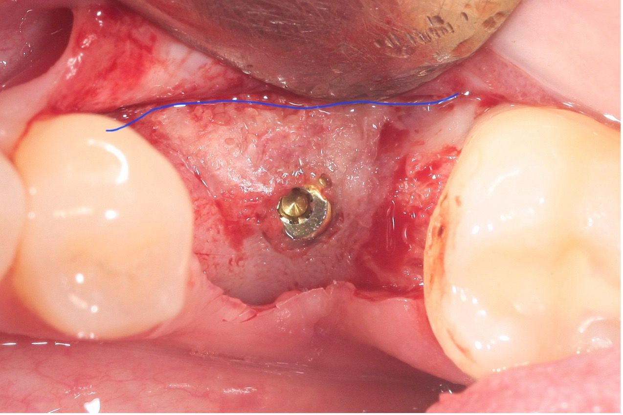

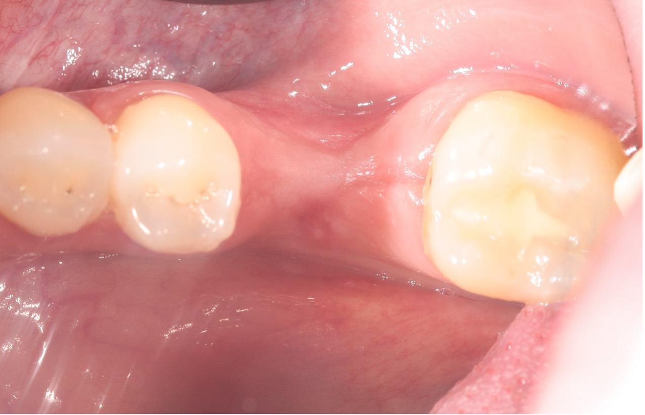
Pre Op - thin alveolar ridge
A clinical view demonstrating a thin alveolar ridge in the area of tooth 36. The ridge width appeared insufficient for conventional implant placement without augmentation.

Implant Placement in a Thin Alveolar Ridge
Intraoperative view showing implant placement within a narrow alveolar ridge. Despite the limited horizontal bone dimension, the implant was inserted with appropriate angulation and achieved primary stability. Note the prepared site and the positioning of the implant, with clear exposure for simultaneous grafting to optimize long-term support and integration.
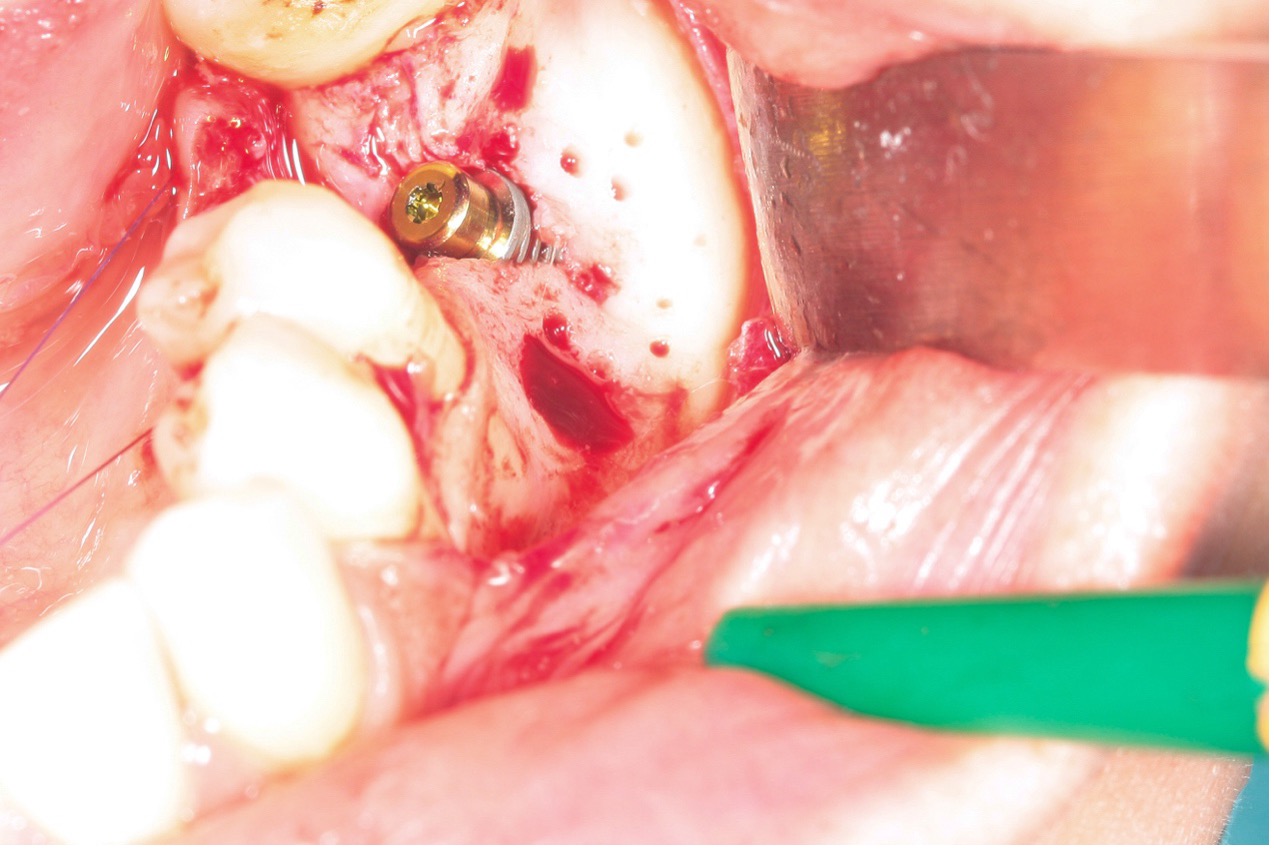
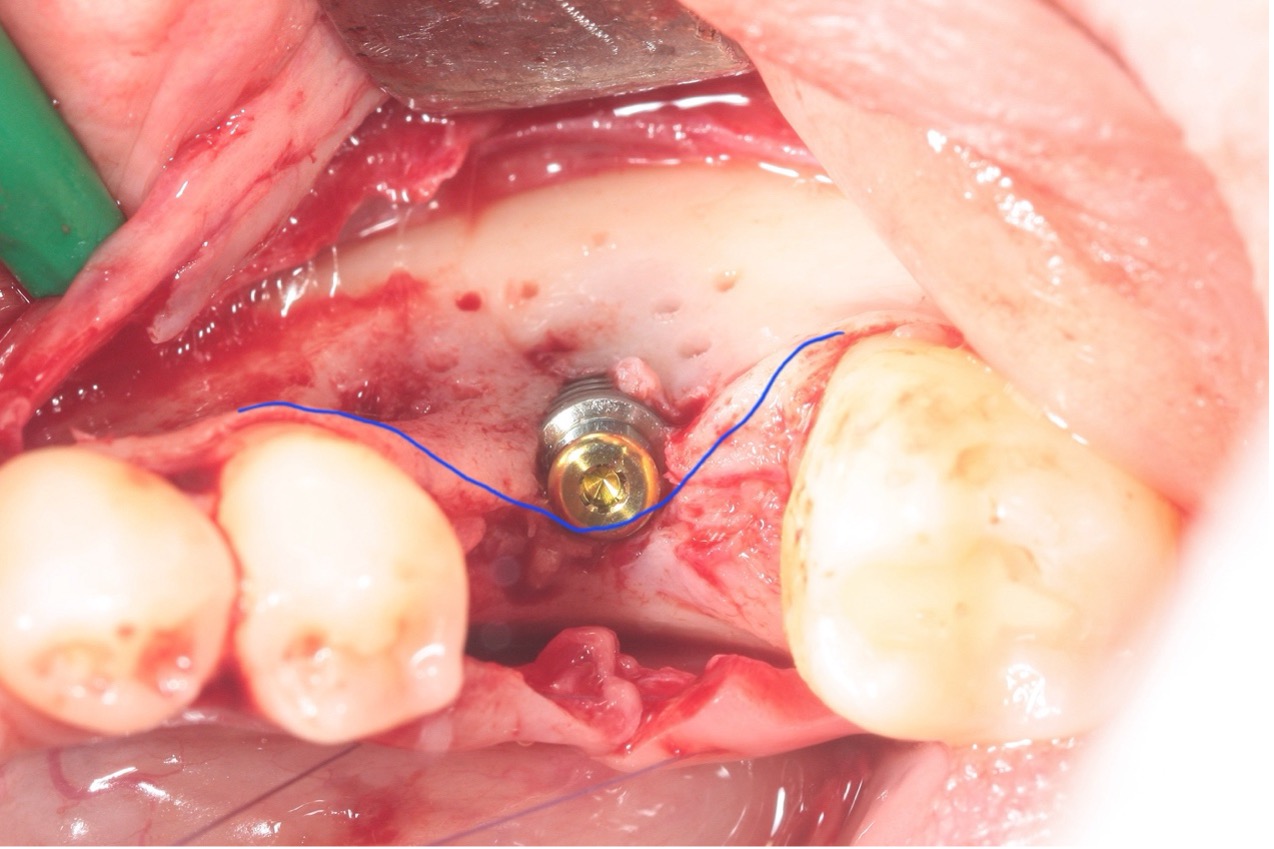
The buccal view – a thin alveolar ridge with exposed implant threads
This clinical image presents a buccal view following implant placement in a narrow ridge. The alveolar bone appears deficient in horizontal width, exposing part of the implant threads. This condition underscores the necessity of simultaneous lateral bone augmentation to achieve optimal long-term stability and full implant coverage. The preparation of the site with cortical perforations is evident, enhancing graft integration and vascular supply.
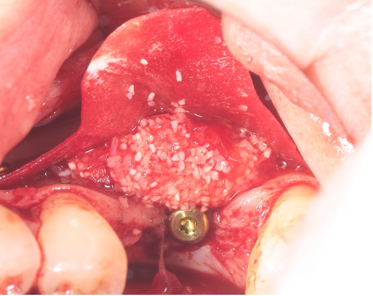
Lateral augmentation using Ivory graft with autogenous bone chips collected from the drills (about 20%)
Grafting phase of the procedure, where a blend of Ivory Dentin Graft and autogenous bone chips—collected from the drilling process—is packed into the lateral defect. The granules are evenly distributed along the buccal aspect of the ridge, ensuring optimal contact with the host bone. This technique enhances regenerative potential while promoting stability around the implant during early healing.
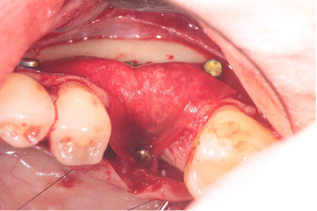
Membrane Fixation – Sausage Technique
a pericardium membrane stabilized over the grafted site using fixation pins, consistent with the “sausage technique.” This approach provides containment of the graft material, promotes space maintenance, and facilitates tension-free closure. The membrane contours smoothly over the ridge, securing the underlying Ivory graft and autogenous bone mixture in place to support optimal bone regeneration.
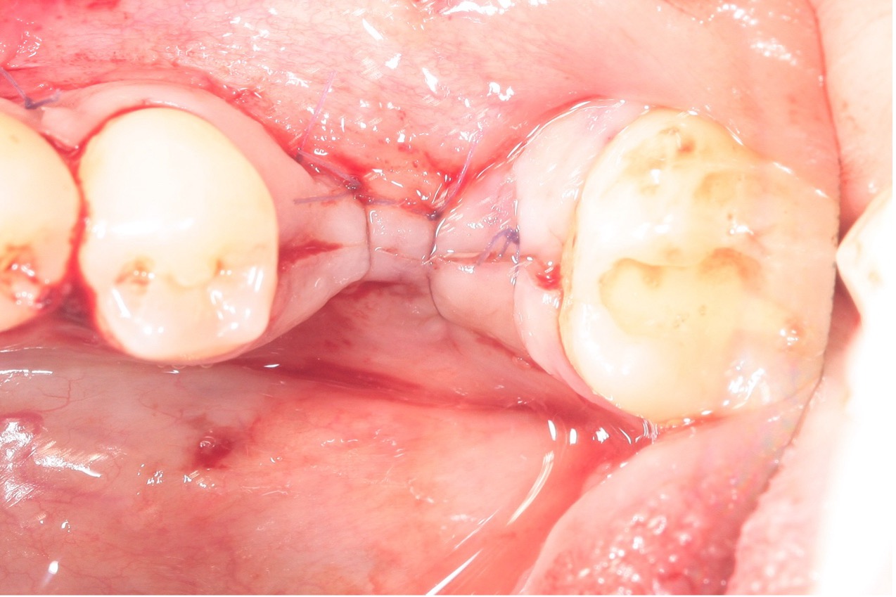
Sutured site
The grafted area is secured with tension-free sutures, ensuring soft tissue coverage and optimal healing conditions.
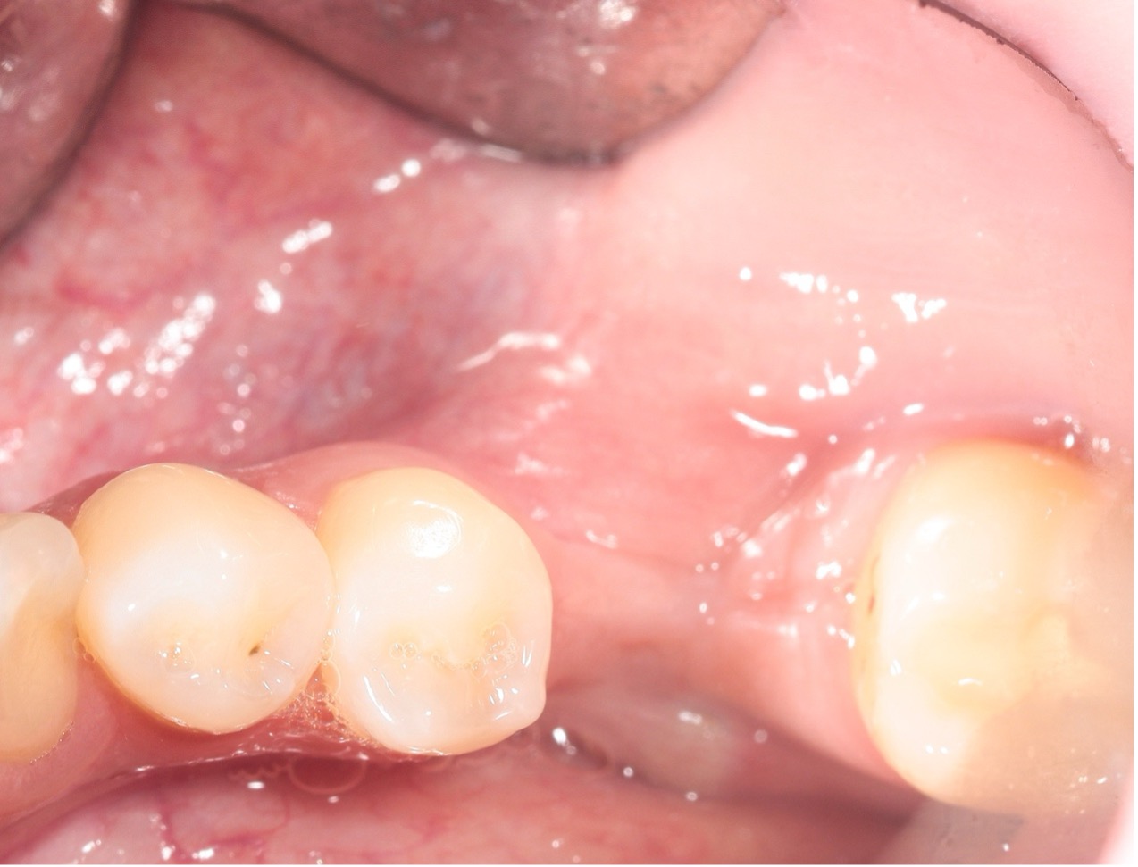
Four months post op
The site shows excellent soft tissue healing with a well-contoured ridge, indicating successful graft integration.

Reopening and Implant Uncovering
Four months post-augmentation, uncovering reveals excellent bone regeneration with dense, well-integrated grafted tissue around the implant. Ridge contour and stability are optimal for prosthetic progression.
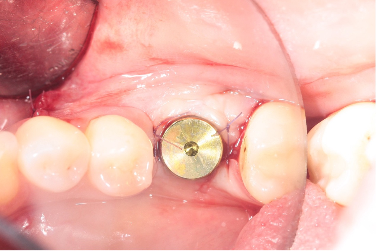
Healing Abutment Placement
Following uncovering, the healing abutment is placed. Soft tissue closure is tension-free, and the surrounding tissue shows favorable healing, supporting a stable emergence profile for the future prosthetic restoration.

comparison pre and post augmentation
A direct comparison of the surgical site before and after grafting demonstrates the clear horizontal bone gain achieved using Ivory graft combined with autogenous bone. The implant threads, previously exposed, are now well-covered, indicating successful augmentation.
Case Outcome:
This case highlights the successful use of Ivory Dentin Graft with autogenous bone in simultaneous implant placement and lateral augmentation. A collagen membrane and proper fixation resulted in excellent regenerative outcomes and implant stability, confirmed at 4 months post-op.


